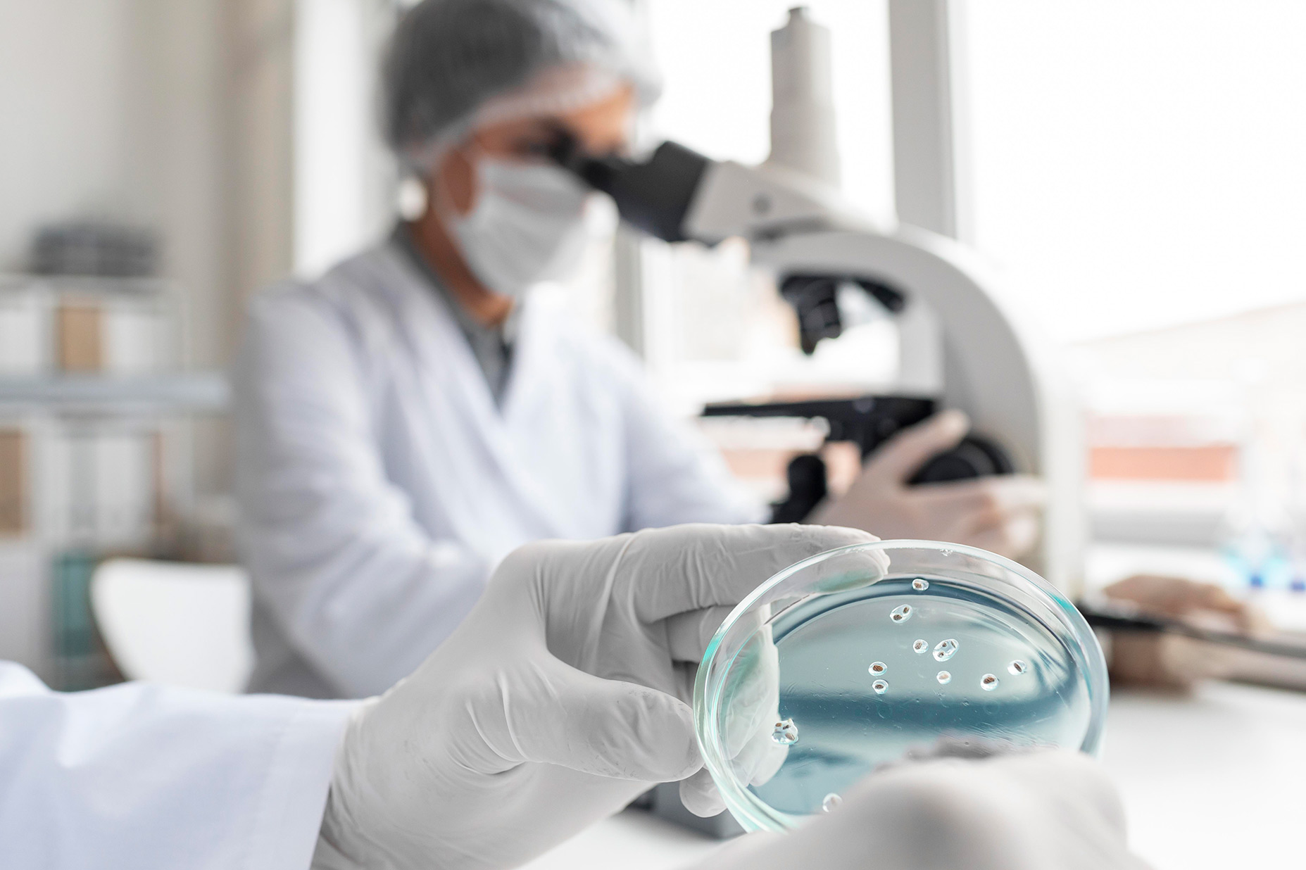
The second year of medical school introduced me to my second favorite subject in the curriculum (second only to Pathology). Knowledge of bacteria, fungi, parasites, and viruses was all tested in the typical one-liner, short-answer, and long-answer questions. As said in a previous entry the written/lecture portion is followed by the practical portion for understanding microbiology. The practical portion consisted of identification (cultures, bacteria, parasites, equipment) and microscopy staining methods (Gram Staining versus Acid Fast staining).
The lectures
Lectures with varied subjects were taught in lecture halls during the morning. The practicals were given in the afternoon with the varied subjects alternating by the day. The microbiology practical started with an introductory PowerPoint presentation taught by an instructor. It included the bacterial staining of that day and spots. The topics ranged from differential media to autoclaves to E. Coli cultures. After the lecture the active practical portion took place to hone in understanding microbiology.
One of the first techniques taught to us was gram staining a pre-made slide following the proper procedure, which I will share with you because I had to memorize it and perform it during my exams. The first step is to apply the crystal violet (primary stain) to the slide. After, a mordant (Gram’s iodine) is added. Following this, the de-colorization process is done with ethanol or acetone. Finally, Safranin (counterstaining) is applied in order to visualize the slide, which is placed under the microscope. Thus, part of the practical portion is creating a slide and then placing that slide under a light microscope. After you have to correctly identify the type of organism. The requirements included a drawing (gram positive or gram negative), the shape (cocci, bacilli, coccobacilli, etc.), and the exact name of the organism (eg.: Staphylococcus Aureus or Neisseria Meningitidis).
The final stain
The final stain was Acid Fast staining for the Mycobacteria with Acid-fast stain. Carbolfuchsin is applied, followed by acid alcohol. The next step is a counterstain with Methylene Blue. Basically, after the slides are prepared an exam of the practical portion deals with an examiner asking the student about the particular slide that you prepared. The examiner can go into the transmission, media used to grow the organism, treatment for the organism, and complications that can occur if someone is not treated properly. They can also ask you questions regarding instruments such as autoclaves, differential media, selective media, and other organisms.
Understanding microbiology further, one of the interesting subtopics that are dealt with includes dealing with rarer organisms that aren’t found in developed countries. Obviously, there are bacteria such as M. Tuberculosis that are more prevalent in India, and hence why they make you learn the acid-fast stain. There are more common infectious diseases spread by Chikungunya, Malaria, and Dengue.
Other rare parasites such as Taenia Solium, Giardia Lamblia, Oochoncerosis, and Taenia Saginata are important to the practical portion in terms of identification and drawing the particular organism because there are cases that come to the hospital. Similar to the gram stain or acid-fast stain, the parasite slide is tested by having the examiner ask for the organism’s Id. Afterward, the examiner can ask you what are the preventive measures (eg: mosquito nets and sprays for Malaria), treatment (including the type of drug and mechanism of action), and life cycle of the organism.


